By John Walsh, PA, CDE and Ruth Roberts, M.A.
Authors of Pumping Insulin and Using Insulin
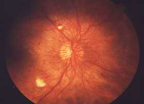
Proliferative Diabetic Retinopathy can be seen in the eye with an ophthalmoscope as neovascularization, a proliferative growth of abnormal new blood vessels. Neovascularization appears as a twisted collection of blood vessels and is quite dangerous because these vessels grow abnormally out of the retina into the clear vitreous gel. This abnormal growth of blood vessels can be seen in the center of this picture. Right above the center is an example of the vessels groeing out of the retina. Because vessels grow beyond the supporting structure of the retina, they are very prone to bleeding, especially when they occur near the disc, the area where blood vessels and nerves enter the eye. Any jerking motion or even a rise in the blood pressure can lead to a rupture of one of these abnormal vessels and cause an hemorrhage.
Bleeding into the vitreous stops the transmission of light into the eye and may be noticed as red, grey, or black blots in the visual field. If bleeding is extensive, a rapid, painless blackening of vision occurs. Later, over a period of weeks, blood slowly clears from the vitreous cavity and vision returns. At that point, an ophthalmologist can start to see the retina and use laser treatments to close off these dangerous blood vessels.
If extensive or repeated bleeding occurs, fibrous tissue or scarring can form on the retina. Since the retina is a thin structure like tissue paper and is made up of only a few layers of cells, scarring can easily pull or detach the retina. Retinal detachment may be noted as wavy lines or a curtain-like effect that appears in one area of vision. These wavy areas are not always obvious, but can sometimes be picked up by testing with an Amsler grid (see the self-test). Further loss of vision may occur if the retina is not reattached.
Before bleeding occurs, someone who has neovascularization will usually not be aware they have these abnormal blood vessels. But they can be detected at an early stage with regular eye exams, and laser treatment at this stage will often prevent bleeding from occurring. Of symptoms that give warning, the appearance of any wavy lines, a spider web, or black lumps in the visual field require an immediate visit to the doctor or ophthalmologist.